|
|
The local student chapter of Materials Research Society (MRS) in the Materials Science and Engineering Department at the University of Tennessee hosted the 2026 edition of the "Science As Art" event. They invited undergraduate and graduate students from STEM departments to contribute artistic presentations of their science. Twenty-one works created by UTK students were submitted to the competition. (Click on the poster to image a higher resolution version.)
The exhibit below presents all submissions, beginning with the first three prize winners.
|
|
|
|
|
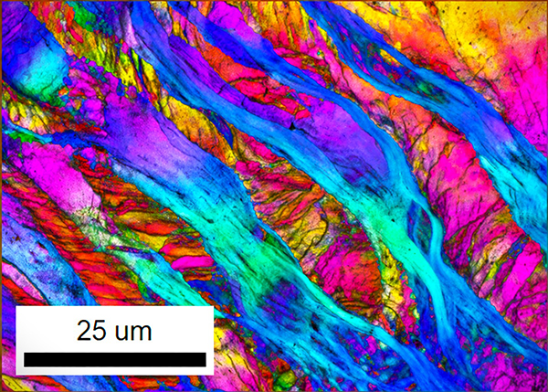
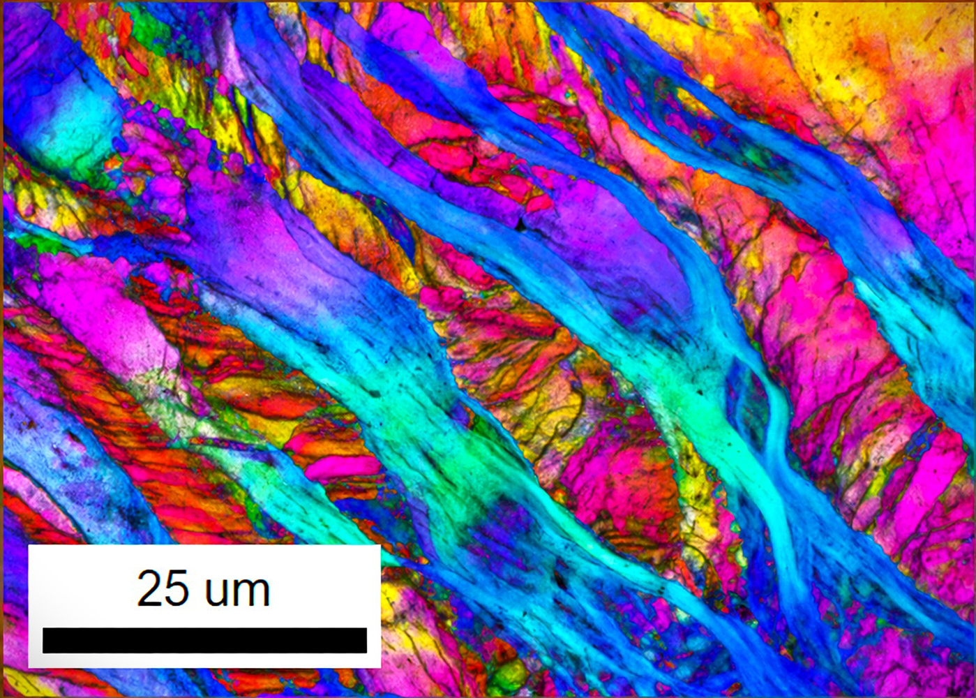
untitled - Joseph Bawah (Materials Science & Eng'g)
"What looks like a painted landscape is a hidden world in motion. Inside high-entropy alloys (HEAs), atoms glide, fold, and bend through dislocation slip, twinning, and kink band formation, leaving behind these flowing traces. Each color marks a path of stress, a memory of force, a moment of transformation. These microscopic patterns show how matter adapts, strengthens, and endures, much like rivers shape the globe. The beauty of materials is revealed in this silent, invisible dance, where structure becomes story, and physics becomes art."
|
|
|
|
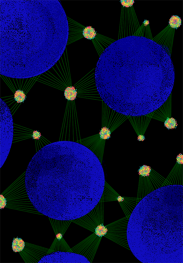
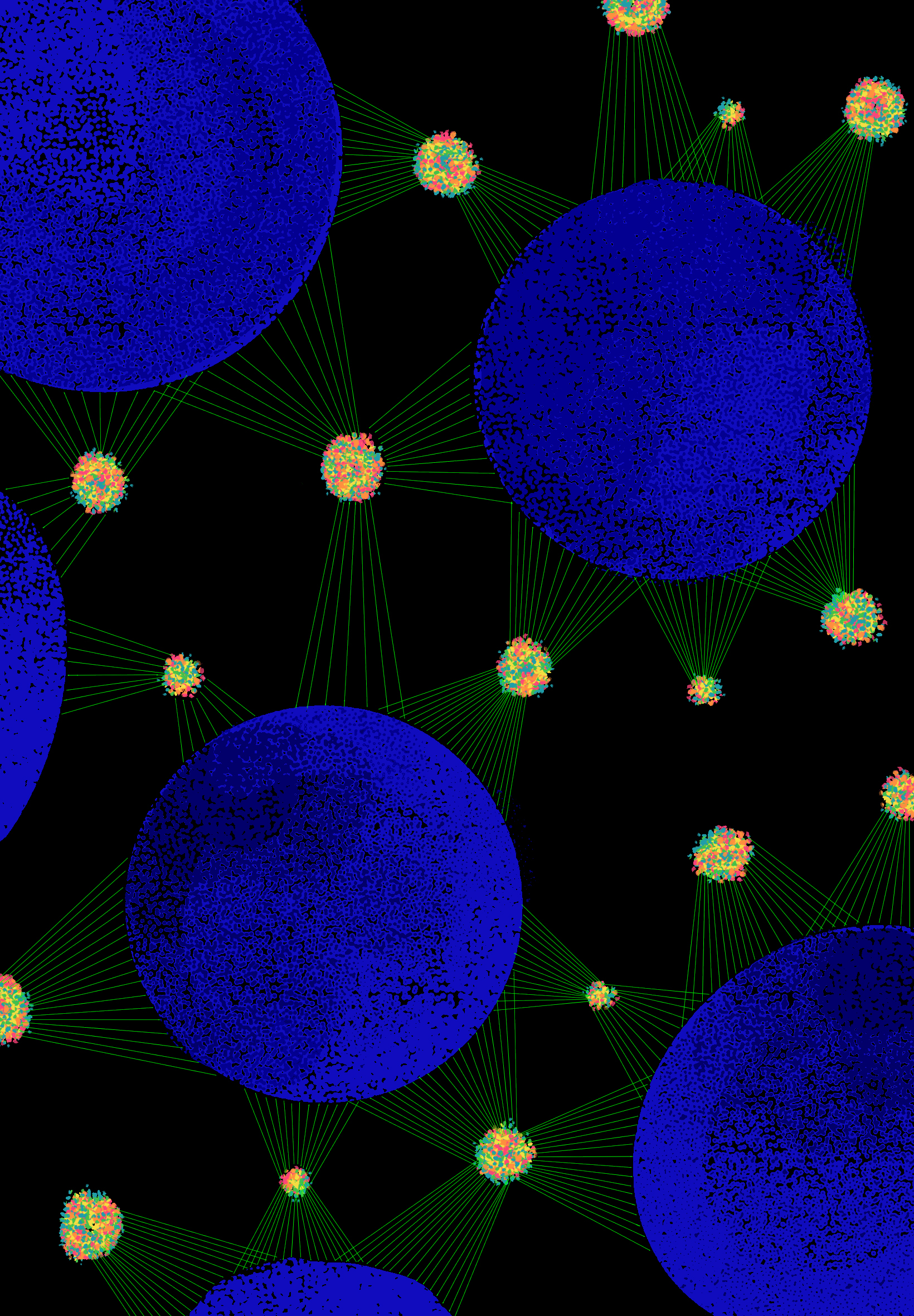
untitled - Emma Morton (Civil & Environmental Eng'g)
This image transforms a microscopic chemical process into a vibrant visual network. The blue spheres represent resin particles, while bright clusters of color show critical metals being captured and held. Thin green pathways connect these elements, illustrating the invisible interactions that guide separation at a tiny scale. Inspired by scientific imaging techniques, the piece blends structure and color to reveal patterns that are normally unseen. By reimagining data as art, the image highlights the beauty within complex systems and invites viewers to see chemistry not just as a process, but as an intricate and dynamic visual landscape.
|
|
|
|
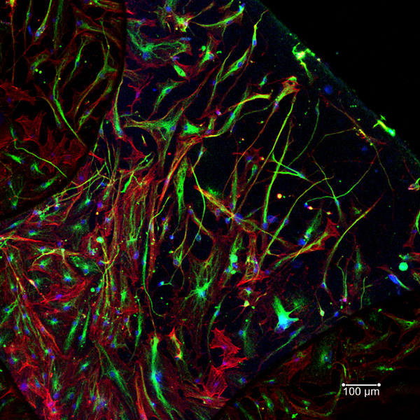
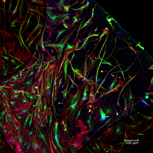
untitled - Eli Christoph (Comparative & Experimental Medicine)
Human stem cells are shown spreading and communicating on a nanoparticle-treated scaffold for regenerative medicine application. The skeletal filaments (red) and attachment proteins (green) illustrate that even at its simplest form, life is defined by beauty, complexity, interactivity, and motion.
|
|
AND THE OTHER AMAZING SUBMISSIONS
(arranged in alphabetical order by artist's last name)
|
|
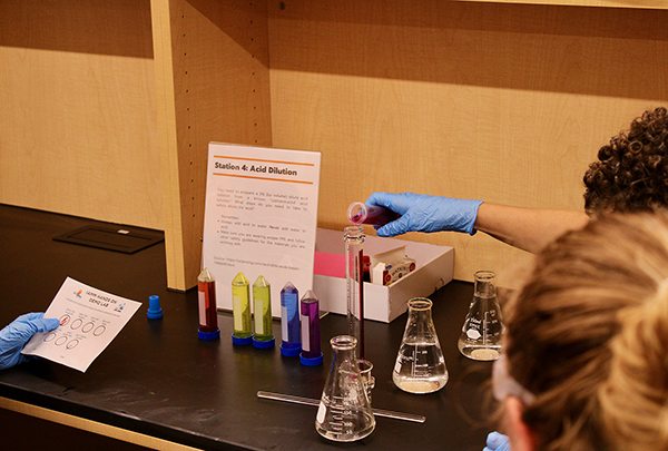
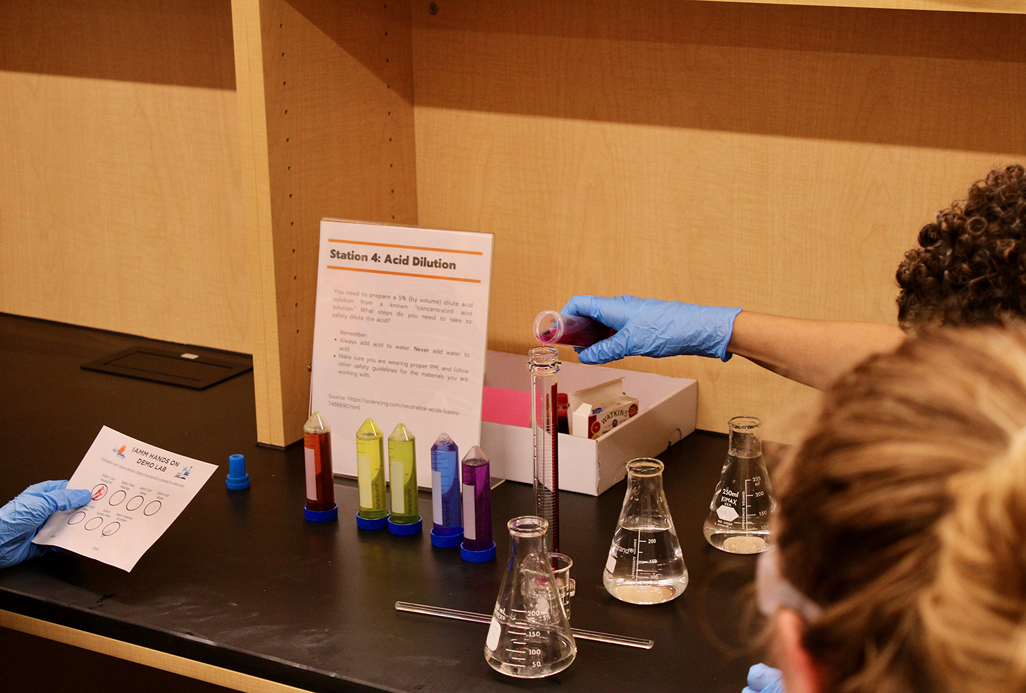
IAMM Lab Safety Day - Courtney Brown (Institute for Advanced Materials & Manufacturing)
Lab Safety Advocates, Grace Loy and Chris Morris, set up a Demonstration Lab in Innovation South last year for folks at the UT Research Park. Students went station to station to learn about how to safely conduct research. In the image, you can see Post Doc, Shelby Watson, watching over Station 4, an Acid Dilution Station. She was teaching students the proper way to dilute acids. You can see her closely watching over the student trying the activity and feel her silent support from the unfocused portion. Another student is watching to the left, waiting in the wings for their turn to try. It demonstrates learning in action and prioritizing safety, while trying something new.
|
|

untitled - Aaron Burns (Civil & Environmental Eng'g)
This photograph captures a pair of honeybees gathering pollen on a passionflower during my field research on feral honeybee colonies in Great Smoky Mountains National Park. The vibrant purple filaments and delicate green structures of the bloom create a striking natural stage, where the bees' intricate movements reveal the hidden rhythms of the forest's pollinator life. While studying how wild honeybees survive without human management, I witnessed moments like this—small, vivid intersections of resilience and beauty. The image reflects both the ecological richness of the Smokies and the artistry found in observing nature up close.
|
|
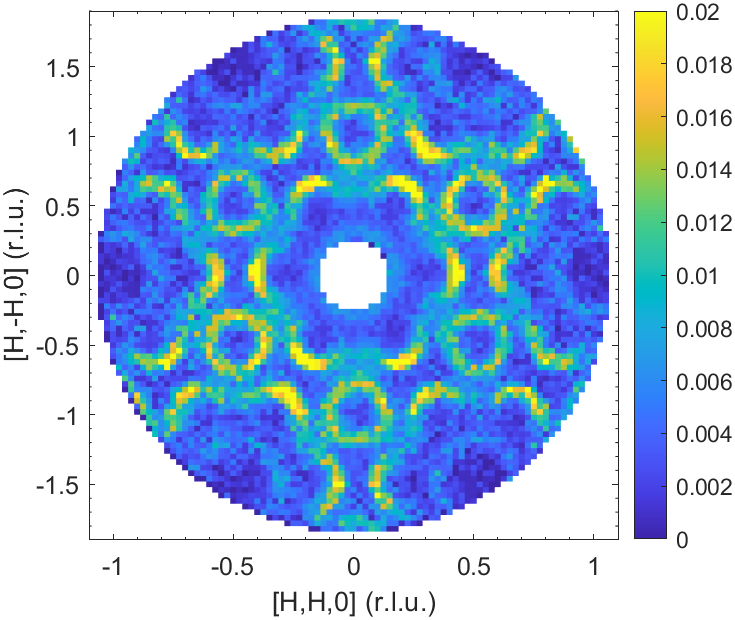
untitled - Emily Chambers (Materials Science & Eng'g)
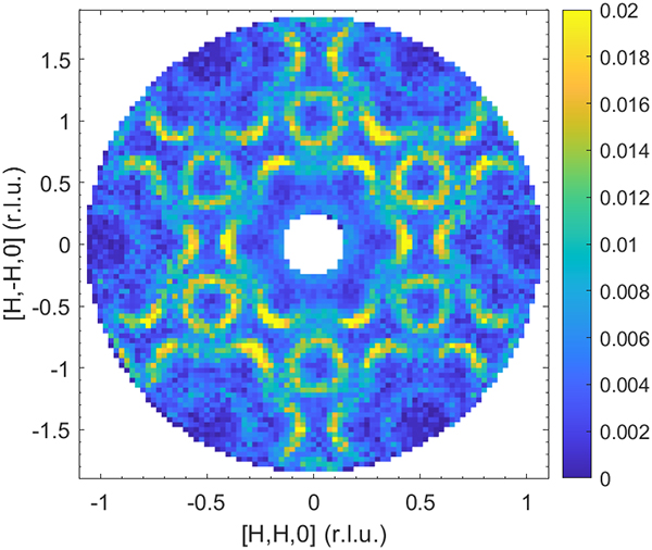
Ce2Zn17 forms a hexagonal crystal structure. Below its magnetic transition temperature of 0.3 K (-273 °C), the unpaired electrons of the cerium atoms form magnetic order. Inelastic neutron scattering was employed at ORNL where neutrons allow us to detect this collective magnetic excitation, which ripples through the material like a wave, thus called a "spin-wave." Here is the low-energy (0.4 meV) spin-wave in the hexagonal basal plane: the sharp, distinct features reflect the magnetic order. Ce2Zn17 is part of a unique class of materials that host unconventional magnetic order, so-called "multipolar order", the origin of which can be challenging to determine.
|
|
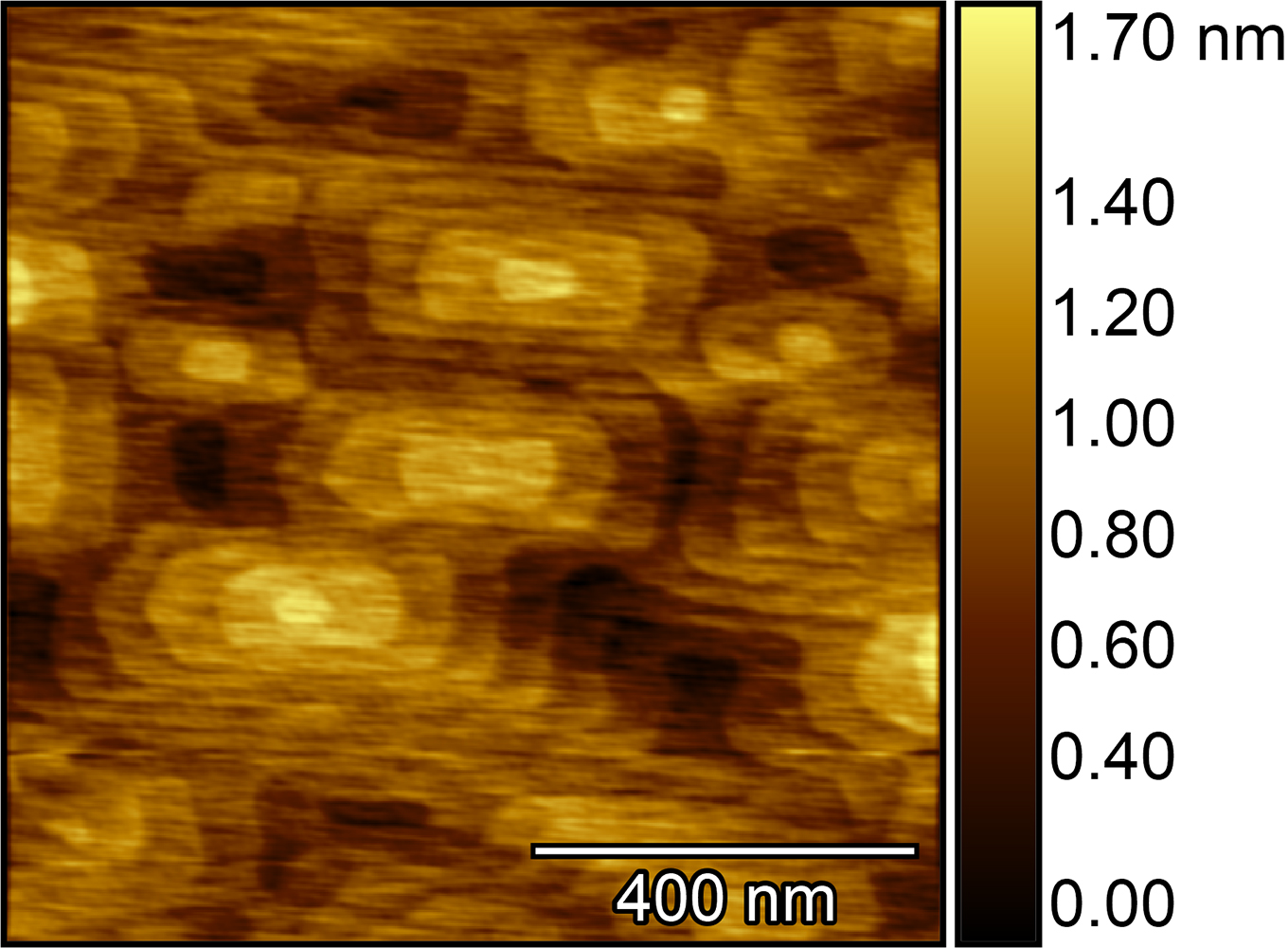
untitled - Chandima Edirisinghe (Physics & Astronomy)
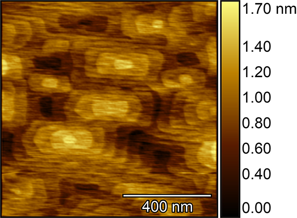
This atomic force microscopy image reveals the surface of a thin film grown layer by layer, similar to a highly controlled form of 3D printing at the atomic scale. The surface appears as a landscape of repeating hill-like structures, where the brightest regions mark the highest points and the darkest areas indicate the lowest valleys. These patterns echo forms found in nature, shaped by repetition and balance. The image's artistic value lies in transforming a hidden microscopic surface into a visually engaging terrain, revealing unexpected beauty and rhythm.
|
|
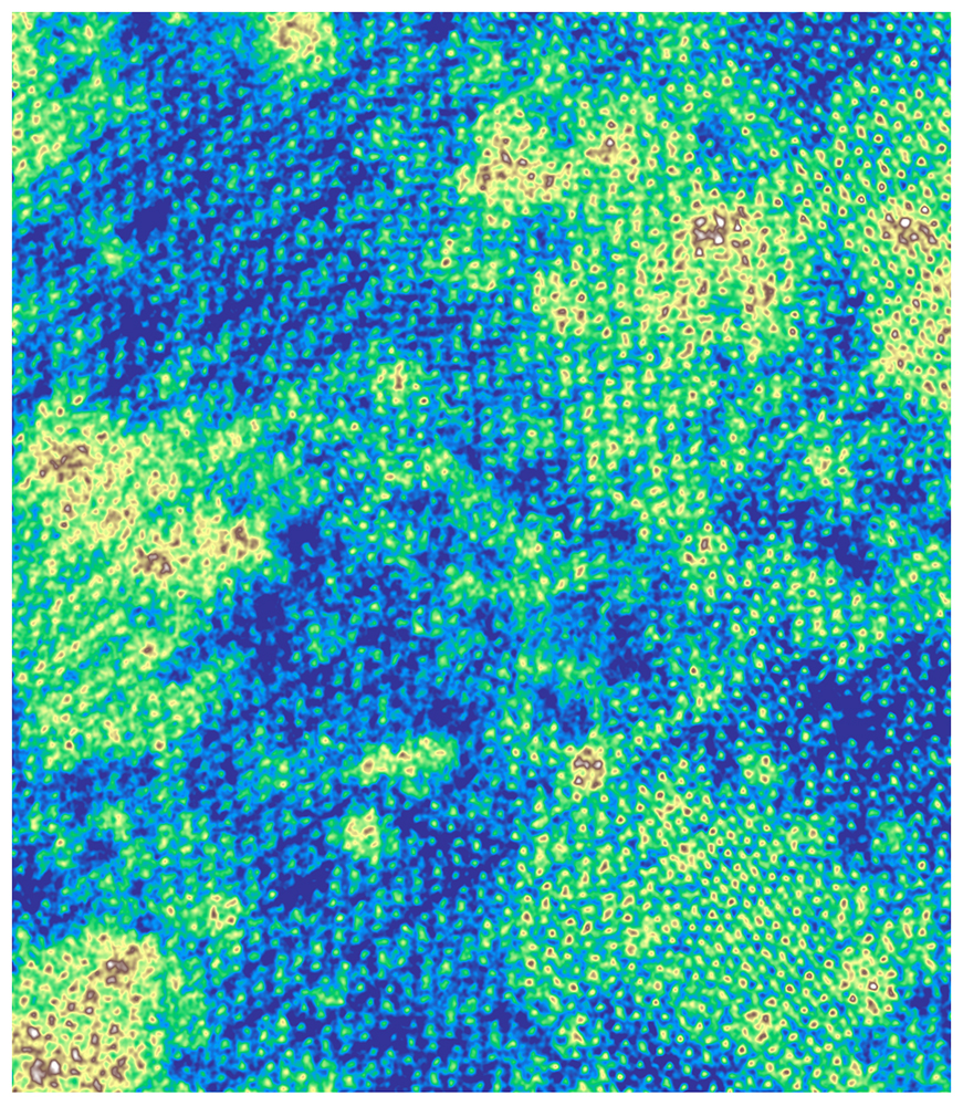
Sailing Seas of Selenium - Austin Houston (Materials Science & Eng'g)
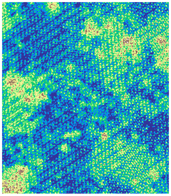
This image captures a monolayer of tungsten ditelluride (WTe2) reshaped by the implantation of selenium atoms. Each bright spot represents a single atom, resolved and mapped with atomic precision. Deep blue regions reveal where selenium has embedded in the lattice, transforming the material's structure. The resulting patterns resemble oceans and continents, yet exist at the smallest scales imaginable. This work explores how matter can be navigated, engineered, and understood—revealing hidden landscapes where new materials and discoveries begin.
|
|
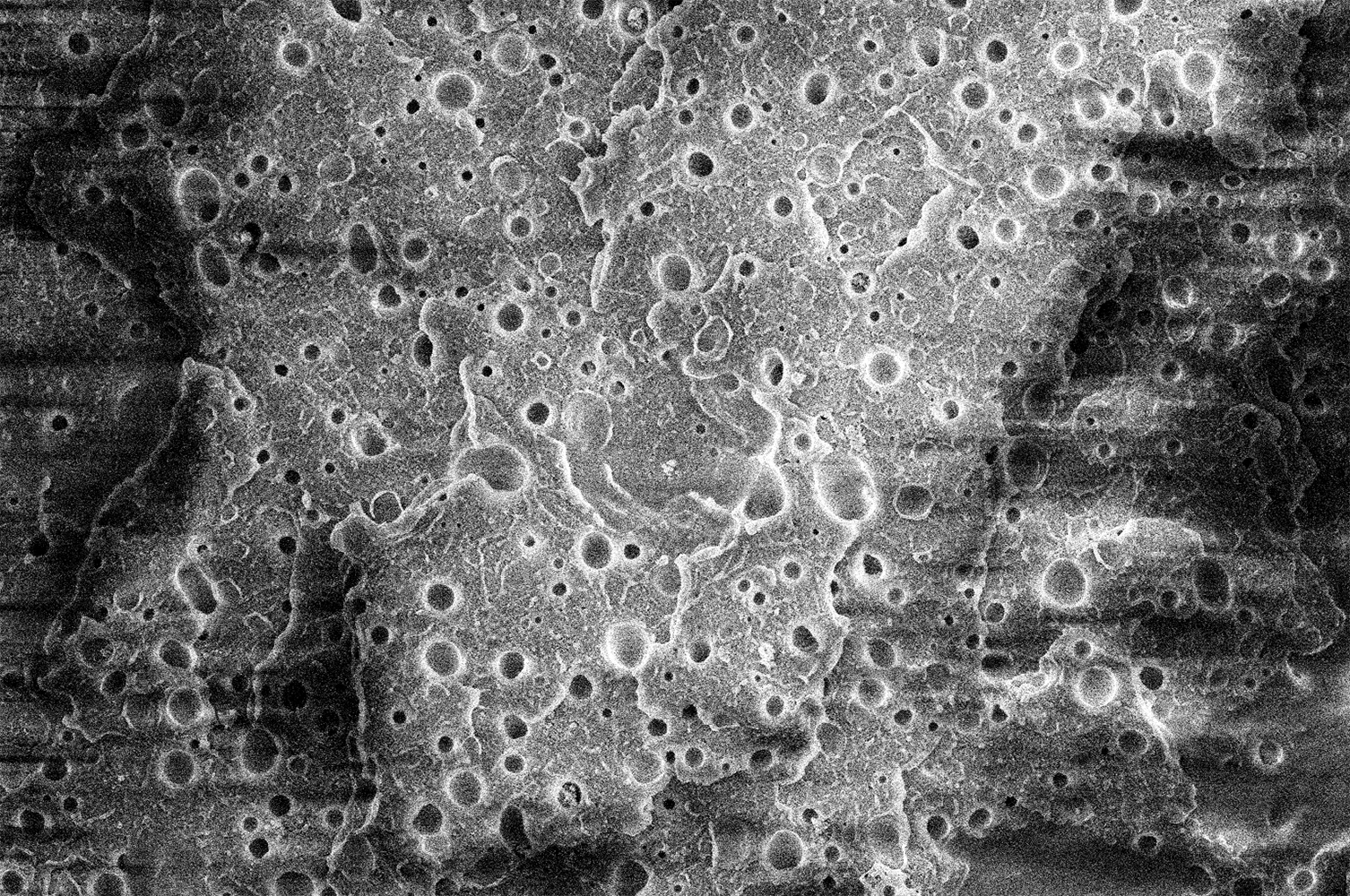
Looking at the Moon - Susan Houston (Chemistry)
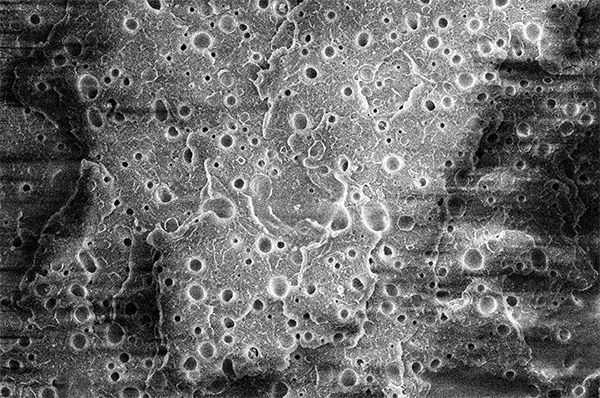
Upon first glance, you may think you are viewing a photo of the moon. In fact, this is an image of the solvent-etched surface of an immiscible polymer blend, where the 'craters' serve as the domains of the minor component of the blend. The whole image is about 25 microns in length. This begs the question: is it really that different to be micro- versus macro-scale?
|
|
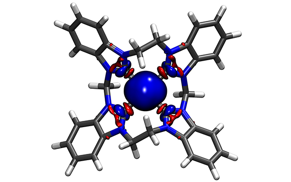
Forging the Bond - Thomas Jones (Chemistry)
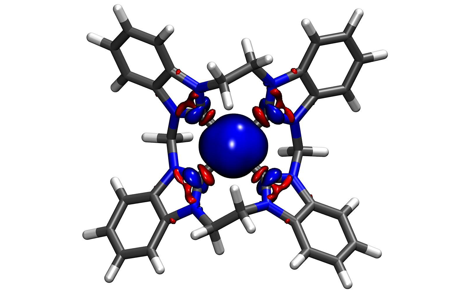
Understanding chemical bonds between transition metals and organic ligands is fundamental to the elucidation of properties of molecular complexes. In this image, we have plotted the net effect between the electrons donated by the ligand to the metal and the back-donation from the metal to the ligand. Due to the high symmetry of the molecular structure (synthesized by Henry Brothers from Dr. David Jenkins' lab, Department of Chemistry, University of Tennessee), we were able to obtain a strikingly symmetric visualization of the charge redistribution, where electron donation and back-donation merge into a single, harmonious orbital dialogue between metal and ligand.
|
|
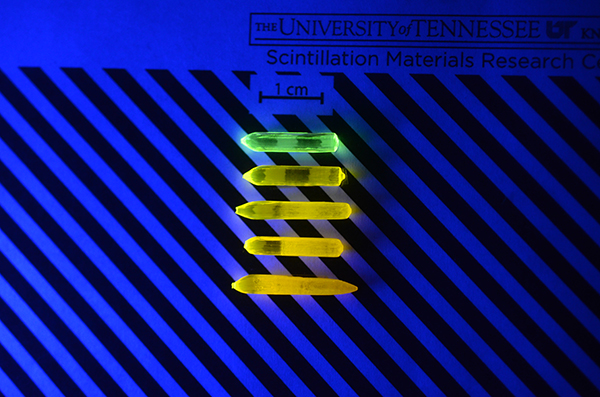
The Alchemy of Energy Transfer - Rebecca Lalk (Materials Science & Engineering)
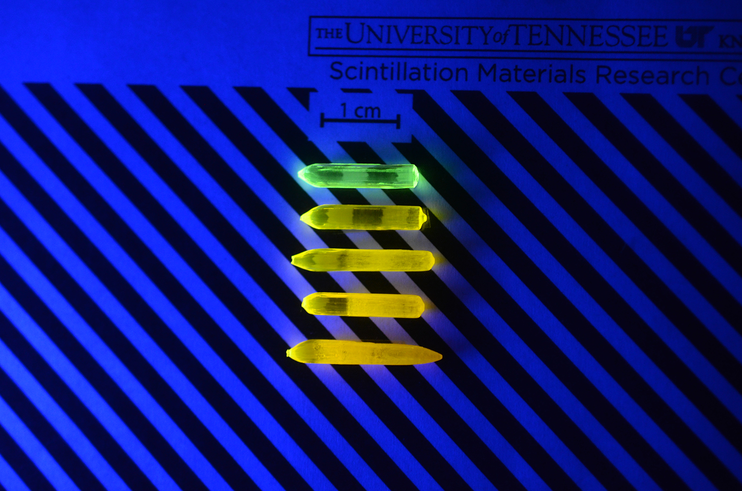
Scintillators are materials that make the invisible visible. When exposed to high-energy radiation, they emit visible light that enables technologies in medical imaging and nuclear security to detect radiation that would otherwise pass unnoticed. Our research develops new scintillators that produce brighter light and faster signals. The crystals shown are (Gd1/4Y1/4Tb1/4Lu1/4)3Al5O12:Ce grown at the Scintillation Materials Research Center at UTK. Under ultraviolet light, the undoped crystal at the top glows green due to terbium (Tb3+). Moving downward, increasing cerium (Ce3+) transfers energy from Tb to Ce, shifting the emission to yellow. Like modern-day alchemy, energy moving between atoms transforms green to gold and helps us see the invisible.
|
|
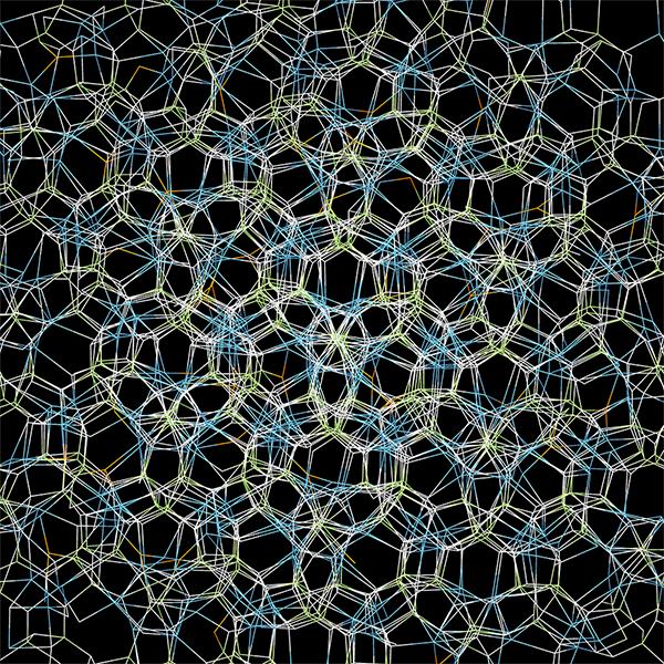
untitled - Katie Loughlin (Materials Science & Engineering)
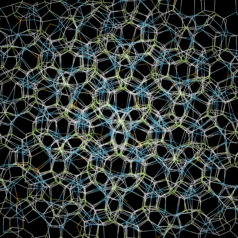
A snapshot of bonds between atoms in a molecular dynamics simulation of Ca12Ga14O33 near room temperature. This cubic crystal, composed of a framework of cages made of calcium (blue), gallium (green) and oxygen (white), also traps other ions (in this case oxygen (orange)) within the cages to balance the overall charge. The image showcases the symmetry of the structure along the body diagonal of the cube.
|
|
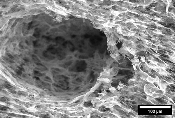
Nanowire Cavern - Maddie Maben (Materials Science & Eng'g)
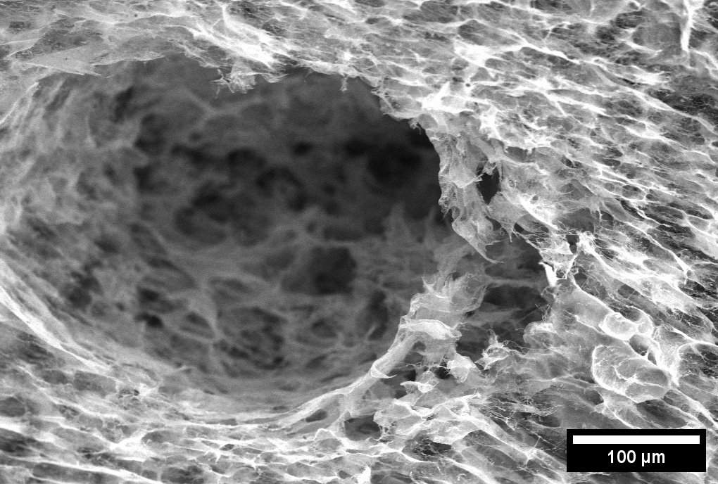
This image reveals a hidden cave within a silver nanowire foam, where delicate strands of metal form a structure that feels both engineered and organic. At its center, a dark hollow opens like a cavern, surrounded by a dense network of shimmering wires that resemble ripples across a quiet lagoon. Though created at the nanoscale, the scene evokes something vast and natural, inviting the viewer to imagine exploring a miniature world. By blending scientific structure with visual intrigue, this image highlights how advanced materials can also possess unexpected beauty.
|
|
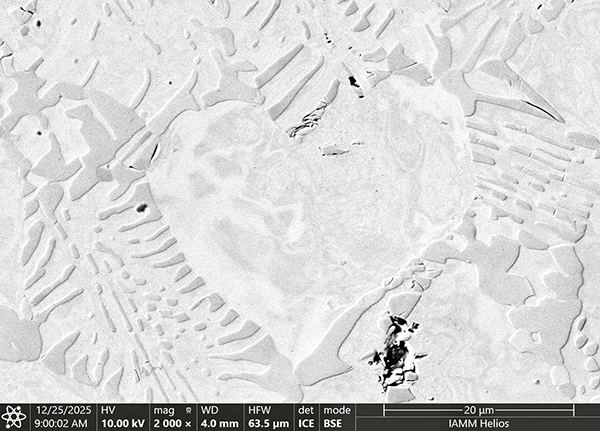
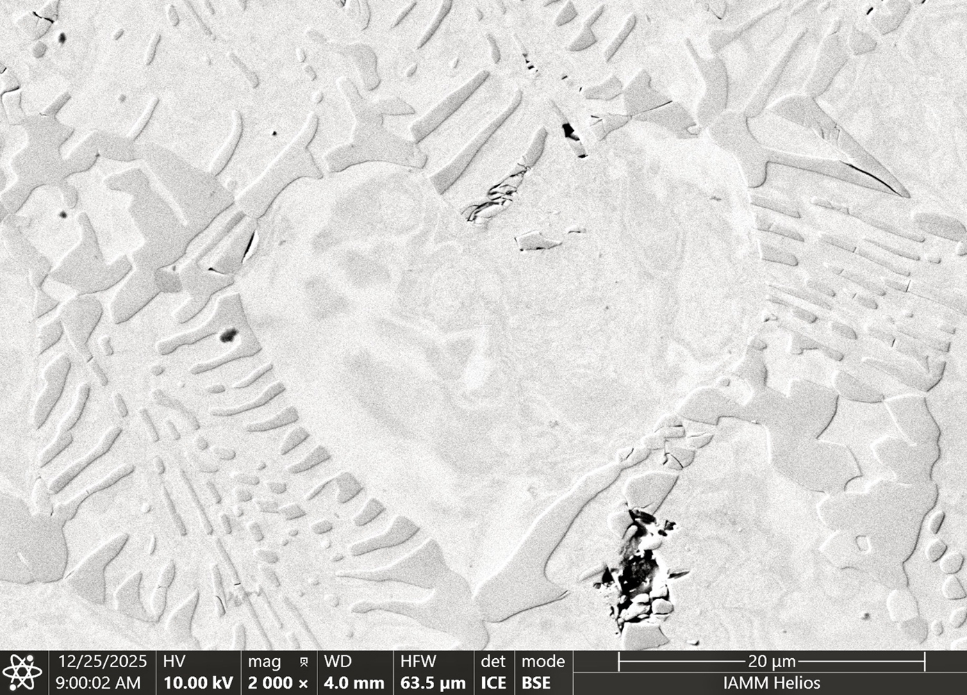
Love is Eutectic - Alfredo Navarrete Nunez (Materials Science & Eng'g)
In our search for creating a new type of Superalloy that can operate at temperatures over 1500 °C (2732 °F) we have encountered some whimsical microstructures that are as intriguing as fascinating. "Love Is Eutectic" comes from an alloy that, due to its composition, forms 2 separate phases instantaneously upon solidification. While studying it with electron microscopy, I felt as if the alloy was observing me. Then I noticed it, a solute poor region that solidified in the shape of a heart, showing me that love can be found anywhere, even in the smallest and most unexpected places.
|
|
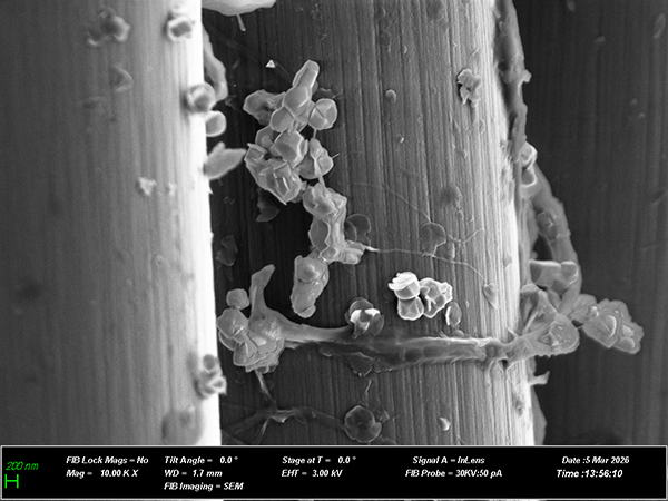
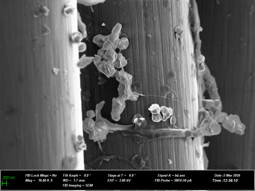
untitled - Adwoa Kyeiwaa Owusu (Mechanical & Aerospace Eng'g)
This image may resemble a castle wall draped with delicate vines and blooming flowers. While it appears artistic, it actually reveals the hidden architecture of an advanced composite material. Composites are made by combining fibers with a surrounding polymer, and their performance depends greatly on how well these components bond together. In this work, the fiber surface—our "castle wall"—is coated with a cellulose-based layer that carries tiny ceramic particles. These particles, the "flowers," give the material a remarkable ability: converting mechanical stress into electricity and vice versa. Beneath this imagined garden lies a glimpse of how scientists design advanced materials that multifunctional.
|
|
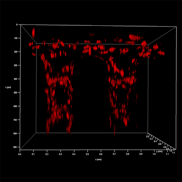
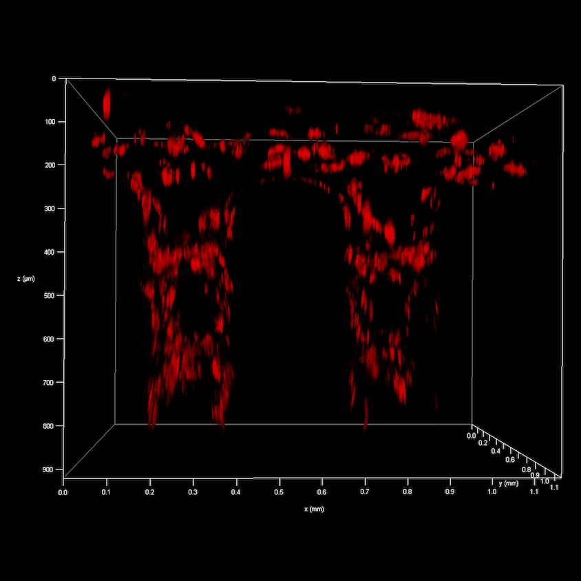
untitled - Berfu Ozmen (Genome Science & Technology)
This image captures labeled stem cell integration into a 3D-printed, grid-designed scaffold for regenerative therapies. The scaffold mimics tissue structure and function, while the cells promote healing. Together, they support tissue regeneration. By labeling the cells, we can track their response to our materials and visualize them within the 3D space of the scaffolds. This demonstrates that we can effectively deliver our stem cells to injury sites using scaffolds and achieve our goal: to accelerate repair and help patients in the clinic.
|
|
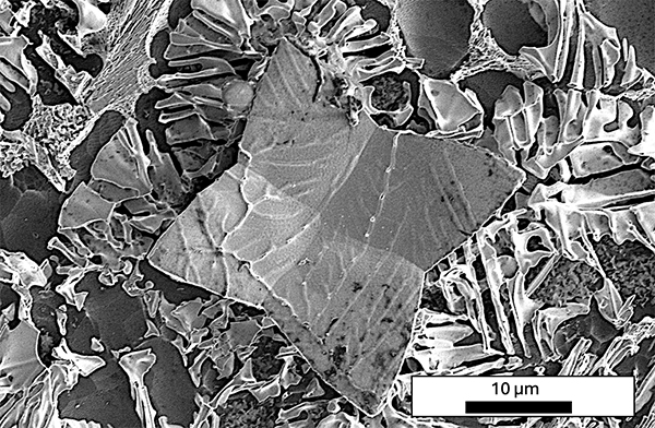
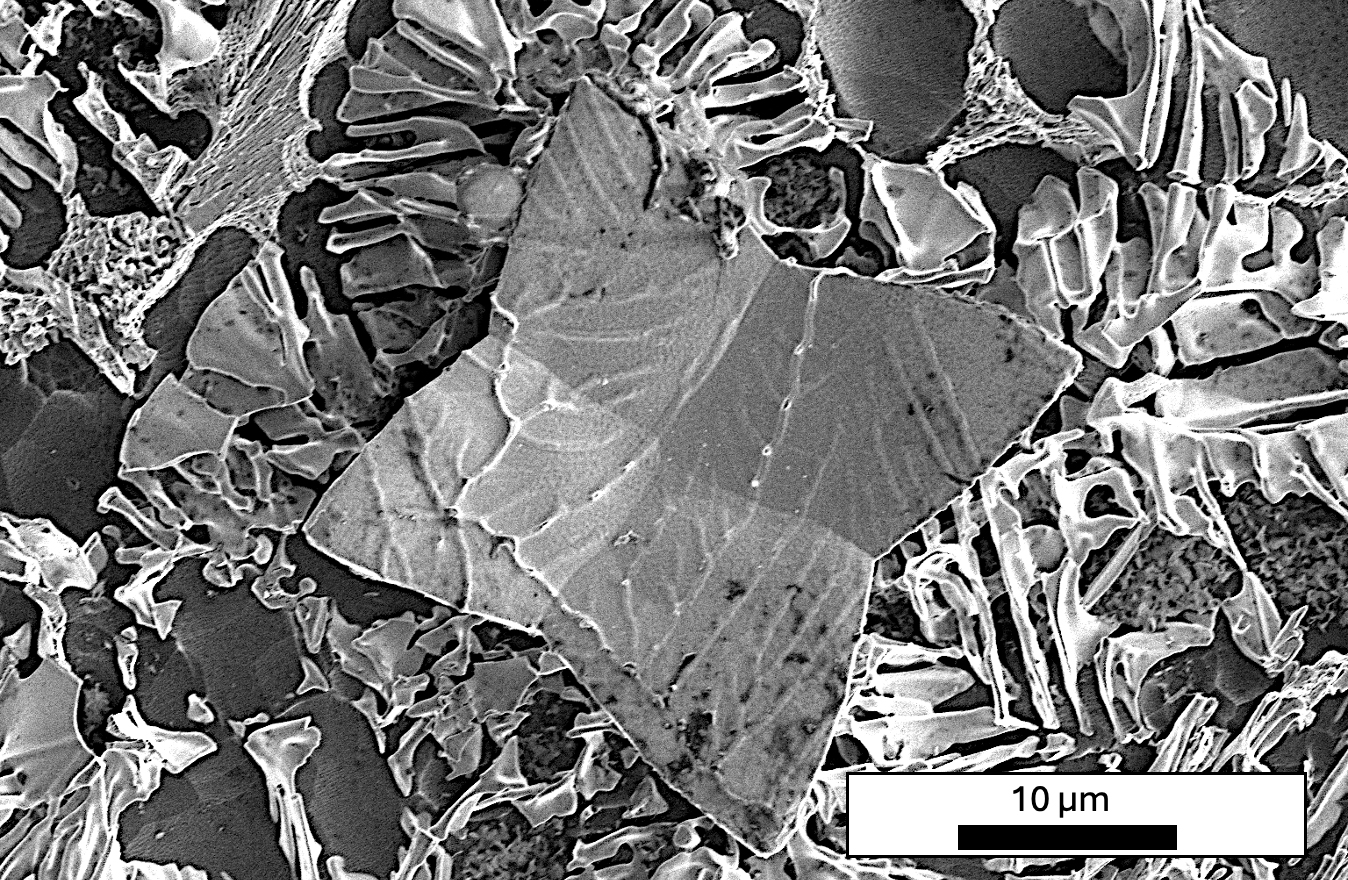
Butterfly Cube - Darel Pates (Materials Science & Eng'g)
This image is of an acid etched aluminum alloy crystal and the mixed metal matrix growing out from the the crystal arms. This will demonstrate a 3 dimensional relationship between the primary crystals that form during casting and the matrix that grows and distributes strain through the material, giving a better understanding of elastic properties.
|
|
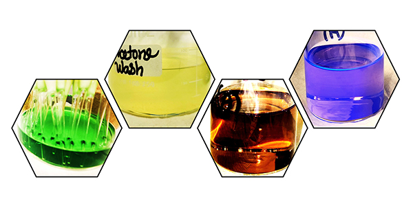
Living Palette of Polymer - Farhana Sultana Shanta (Bredesen Center)
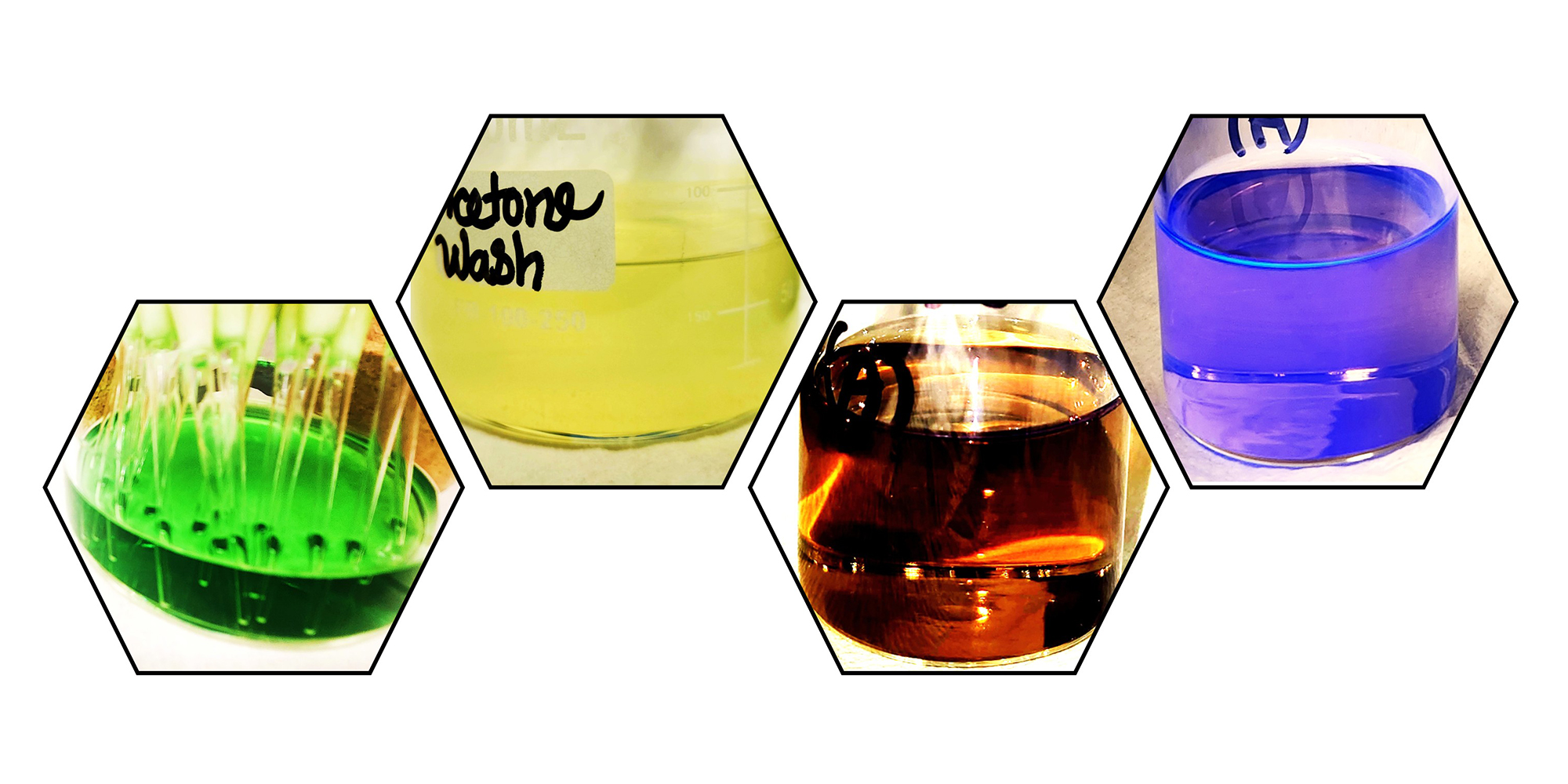
A poem of color written with deep green, soft yellow, dark orange and fluorescent blue is observed during mechanochemical polyfluorene synthesis. A visual journey of polyfluorene from crude state to isolated state (from left to right) transforming the laboratory into a small chromic universe. The fluorescent blue color of purified polymer solution under UV light contrasts with the deep green color of reaction crude mixture in methanol, soft yellow color of oligomers in acetone and dark orange color of isolated polymer in chloroform. An elegant interplay of solvent, purity, and molecular structure, rendered chemistry-as-art.
|
|
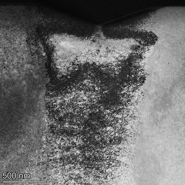
Brittle Butterfly - Thomas Siggilino (Nuclear Eng'g)
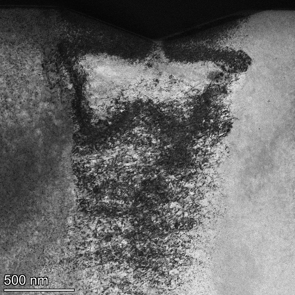
Small defects produced from a combination of deformation and irradiation in a metal alloy form a butterfly shape with a plume of scattered defects trailing under its wings when viewed under an electron microscope. The butterfly also divides light from dark regions as the deformation responsible for its appearance darkens the metal crystal. The strikingly symmetrical butterfly shape arises from a tangle of defects that otherwise appear chaotic, illustrating how order can spontaneously arise from complex physical phenomena. Understanding this complex relationship between irradiation defects and deformation in metals can help design more radiation resistant materials for advanced nuclear reactors.
|
|
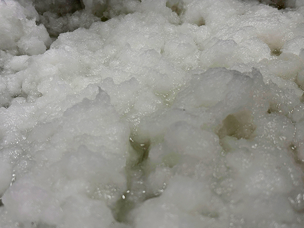
Paper Clouds - Matthew Valderrama (Center for Renewable Carbon)
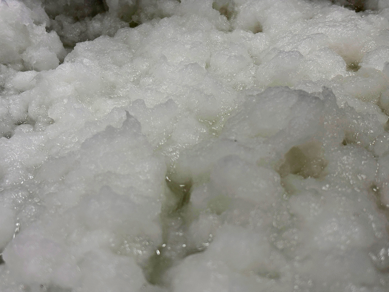
Pictured is a close-up image of a pulp made from paper and polylactic acid fibers. This pulp is a step in my project developing biodegradable and bioderived food packaging. In taking this picture, I wanted to reflect on how paper making is a tradition which has spanned millennia but is still being developed for new applications in materials development. In the same way people can see shapes and figures in real clouds, I see a more sustainable future in my "Paper Clouds".
|
|
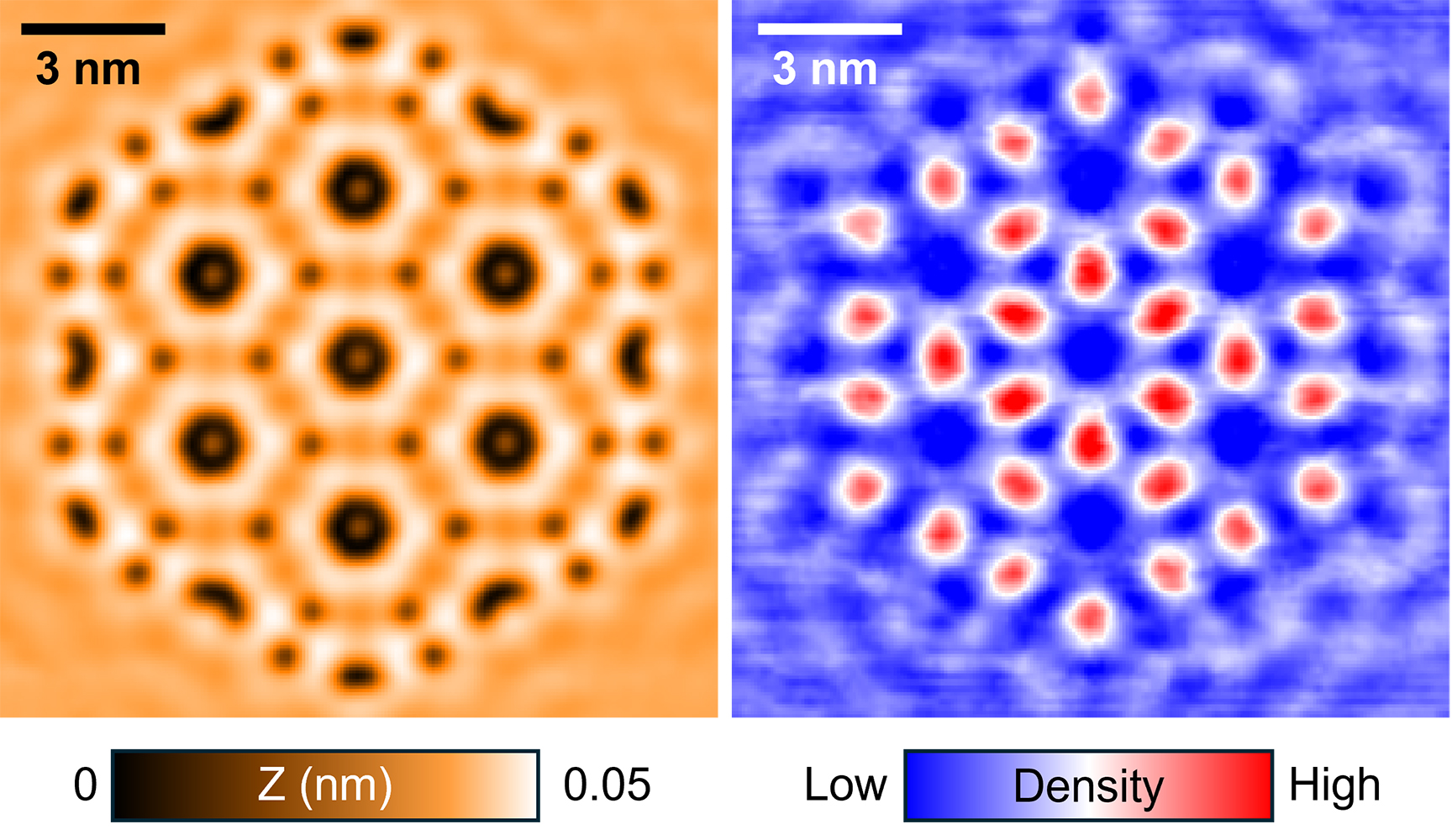
Electrons in a Cage - Wooin Yang (Physics & Astronomy)
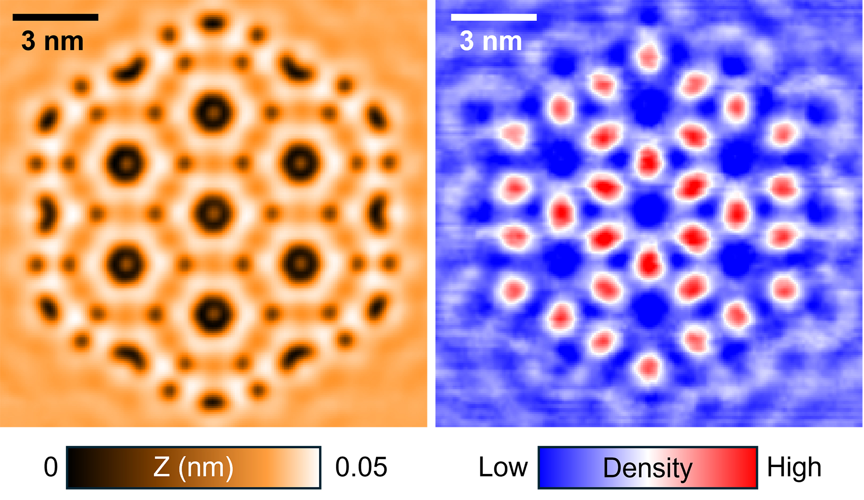
Electrons inside a solid normally roam freely — like ripples spreading across water, dispersive and unconfined. But a well-designed structure can trap them. Using scanning tunneling microscopy, we realized atomically precisely engineered cage (left). The right image reveals the trapped electrons within the constructed architecture.
|
|
|
|
Special thanks to the MSE graduate students who organized this event, especially Elizabeth Heon.
|
|
|
|
More photos on the Keffer research group site.
|
|
|