|
|
The local student chapter of Materials Research Society (MRS) in the Materials Science and Engineering Department at the University of Tennessee hosted the 2023 edition of the "Science As Art" event. They invited undergraduate and graduate students from STEM departments to contribute artistic presentations of their science. Six of these works were exhibited in Hodges Library over the fall semester, 2023. Eleven works created by UTK students are showcased below. (Click on the poster to see a higher resolution version.)
|
|
|

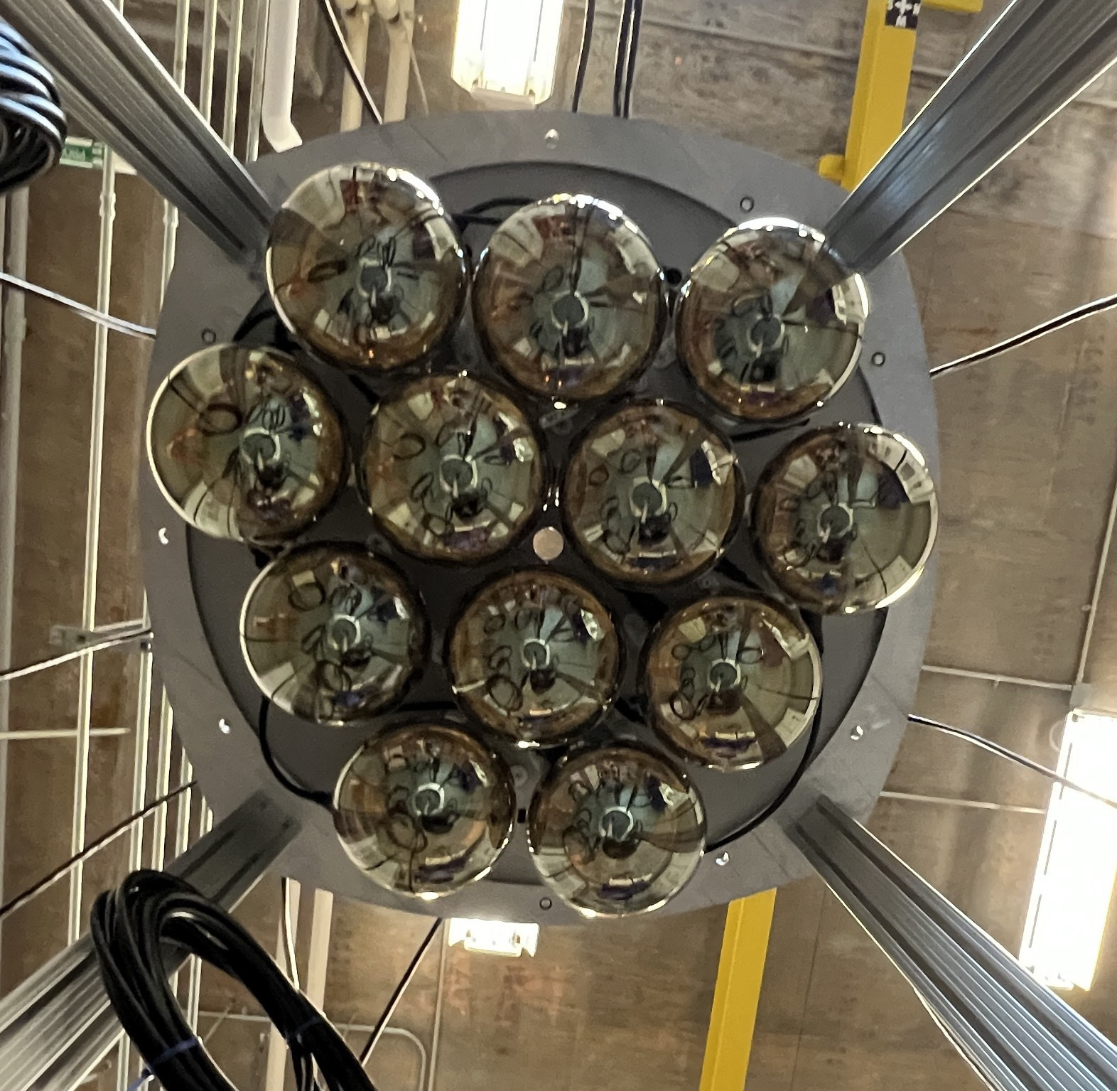
Collecting Light - Igor Bernardi
We usually think of light as what we see. But how can we train a machine to "see" the light? These 12 photomultiplier tubes ("PMTs" for short) may look like light bulbs, but they do quite the opposite: They collect light and generate a signal that can show up on a computer screen. They are essential technological devices for ultra-dark situations where collecting and quantifying the smallest bits of light (photons) are the only ways for researchers to know what is happening on the inside of a detector.
|
|
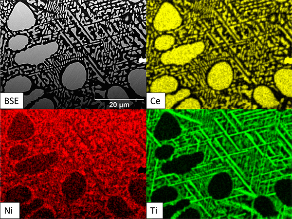
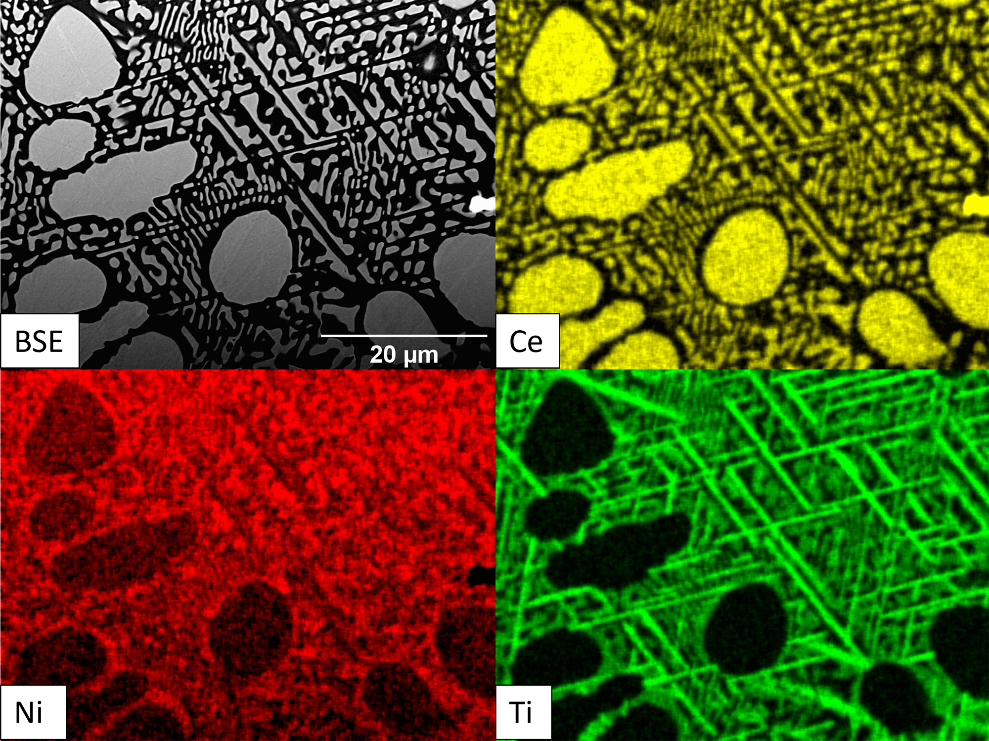
Adding Titanium...Lucky or unlucky? - Syeda Bushra Haider
Different compositions (binary, ternary and quaternary) of Ni-Ce eutectic alloys are currently being studied in the Lass-Group of UTK which have impressive properties for high-temperature applications. Characterizing is the fun part where you can add elements of your choice to the binary to see the effects on microstructures. This image shows the back-scattered image and EDS (energy dispersive x-ray spectroscopy) mapping of Ni-Ce-Ti after heat treatment of 24 hours at 900 °C where the elemental distribution can be seen with formation of eta phase in the eutectic which would be detrimental for properties but great candidate as a piece of art.
|
|
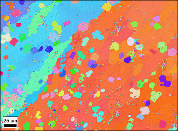
Rainbow Grains - Elizabeth Heon
Most metallic materials are composed of many individual crystals, known as grains. Understanding how large these grains are and how they are shaped is important to understanding the properties of a metallic material. This image was formed by using a technique known as Electron Backscatter Diffraction (EBSD) in which electrons are diffracted, or scattered, off a sample, forming a pattern. The pattern changes depending on how the sample is oriented. Each point is colored based on the orientation of the sample at that point. Because different grains will be oriented differently from each other, we can use this map to visualize the grains.
|
|
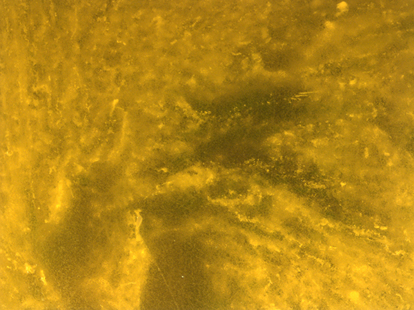
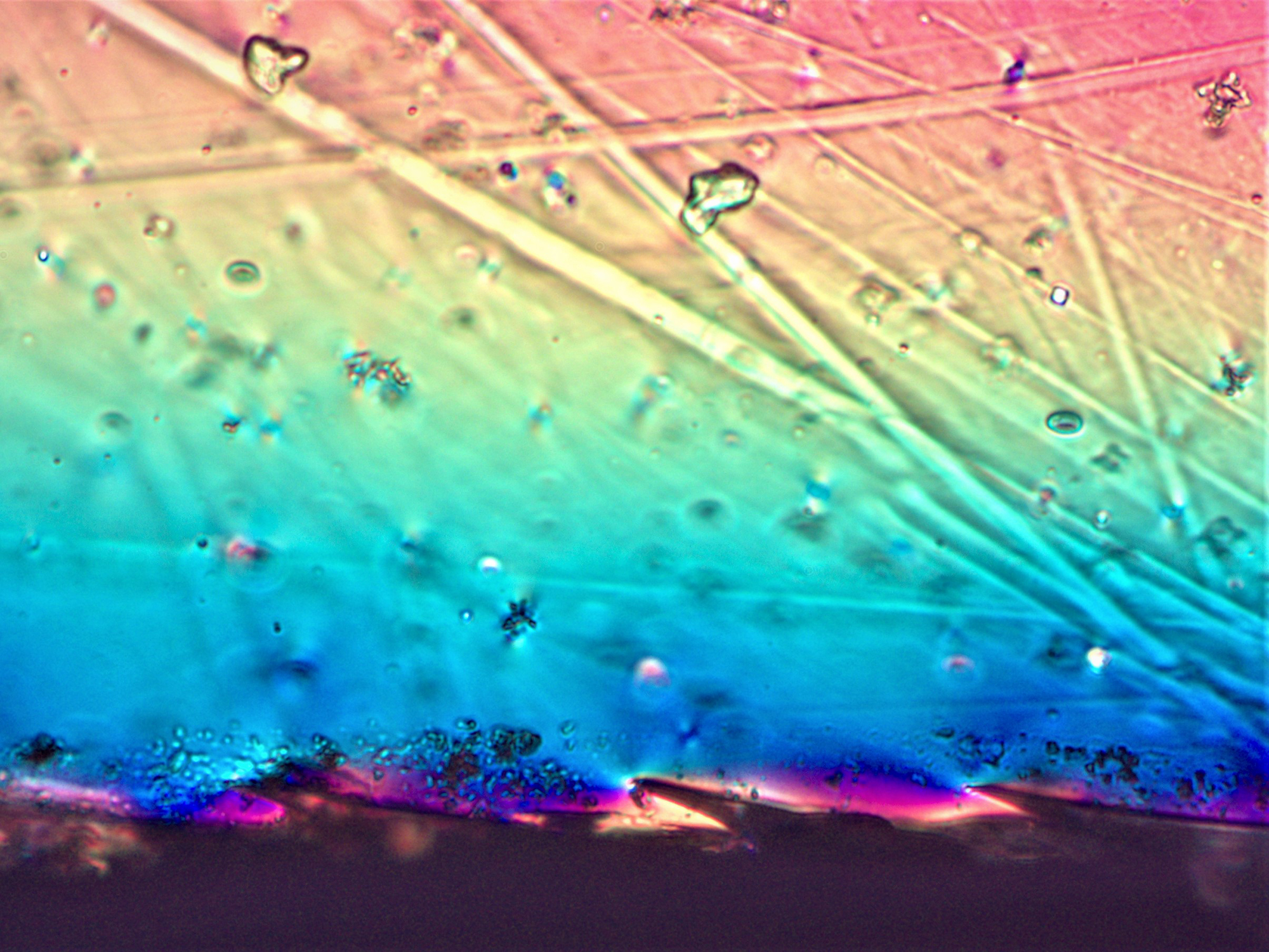
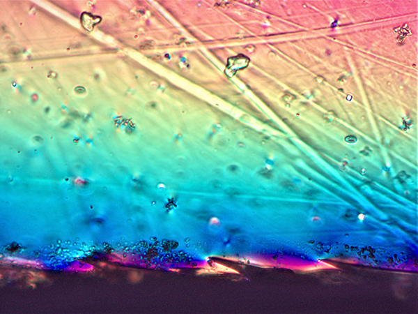
Rising from the Embers - Rebecca Lalk
This polarized light optical microscope image (width = 10 mm) features a cross section of high-entropy aluminum garnet single crystal cut from the tail-end of a boule grown by the Czochralski method. The Czochralski method is a commonly used pull-from-melt technique used in the semiconductor industry. Buoyancy-driven convection, crystal rotation, and melt surface tension drive segregation of certain chemical species, resulting in higher strain and higher concentrations of defects at the tail end of the boule. Cerium doping leads to the crystal's yellow coloring, causing those defects to look like fiery embers.
|
|
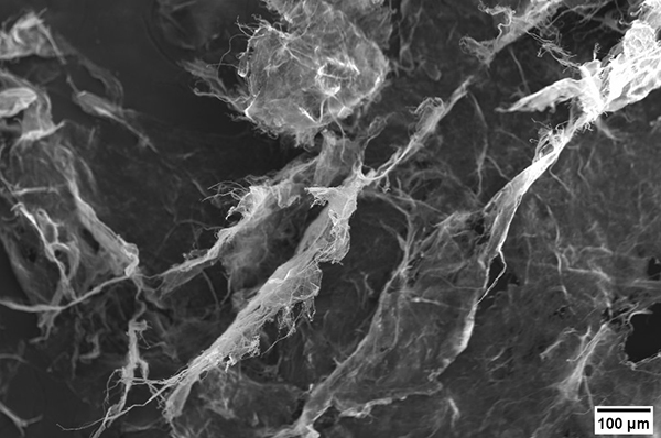
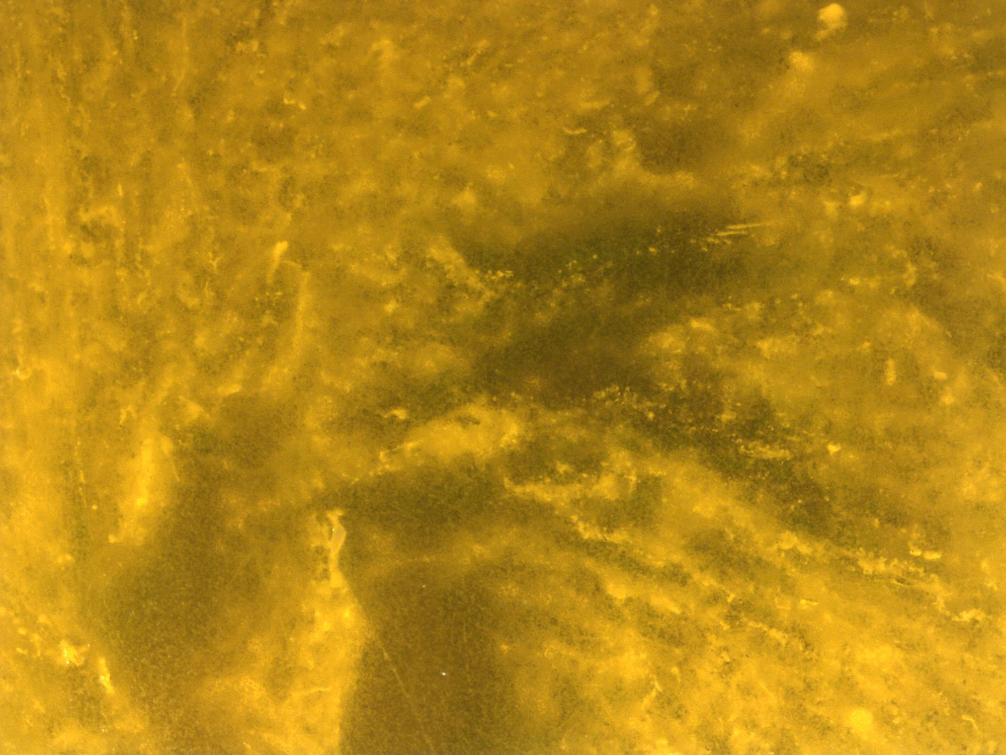
Ghosts of Fibers Past - Melanie Moczadlo
These spooky fibers are nanofibrillated cellulose fibers, which are made by breaking open cellulose fibers using a super mass colloider. These fibers are used in the making of a low carbon footprint, natural fiber composite that is meant to replace current polymers used for interior automobile parts. These nanofibrillated fibers are to help in improving the dispersion of the fibers in the plastic to result in enhanced properties.
|
|
The Prismatic Bridge - Cotton Pekol
Ancient Norse legends tell of a rainbow bridge that stretches from our world to the realms of the gods. This prismatic liquid crystal epoxy bridge synthesized from 4,4'-bis-(2,3-epoxypropoxy)biphenyl and sebacic acid may not reach to the heavens, but it does reach a new world of epoxy functionality. The color gradient in the epoxy bridge indicates crystalline regions capable of shape-shifting and shape-memory activated by heat. With capabilities such as these, this liquid crystal epoxy paves the way for micromachines, self-healing coatings, smart packaging, sensors, and so much more- truly a godsend.
|
|
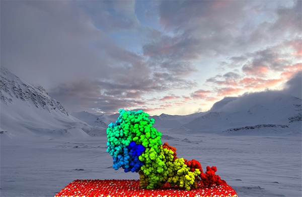
Extracellular Enzymes: The Arctic's Microscopic Polar Bears - Jake Perez
Microbes are not often thought of as keystone species, yet the microbes present in Arctic soils have a strong influence on the polar ecosystem. This polar "bear" is a secreted enzyme, an N-acetyl-β-glucosaminidase, from Svalbard simulated adsorbing to quartz using molecular dynamics. Our research focuses on how adsorption onto soil minerals can impact how these microscopic beasts prey on soil matter and turn it into greenhouse gases, which could further climate change in the Arctic. This first-time sighting, made possible by new bioinformatics tools, will help us understand how Arctic microbes will respond to global warming.
|
|

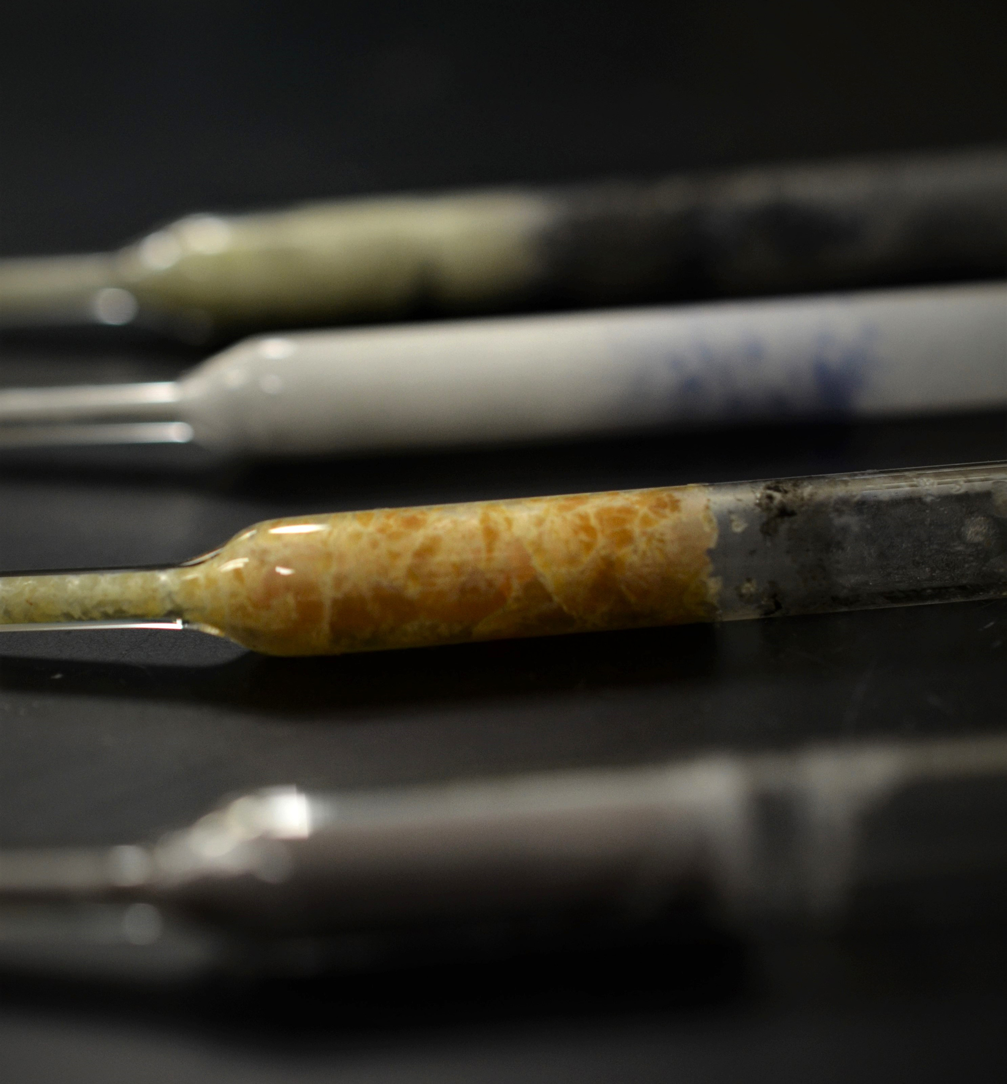
Recognition - Kim Pestovich
Every scientist fails, and that's a good thing. Failure is a crucial part of discovery. However, the valuable results of failed experiments are rarely published. To give failure the recognition it deserves, this image depicts crystal growth experiments that failed for different reasons. The raw materials were poor quality, the compound wasn't stable, or the performance wasn't desirable for the intended application of radiation detection. This knowledge will feed forward into new experiments, accelerating the discovery of new materials. Discovery of new materials for radiation detection can unlock potential of new technologies focused on human health and safety."
|
|
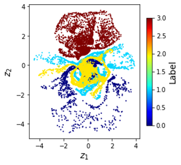
The Hidden Flower - Utkarsh Pratiush
The displayed plot looks like a flower, but it's more than just an aesthetic representation. This is the latent embedding of a machine learning technique called Variational Autoencoder (VAE) trained on card's data to discern patterns. In the field of material science, this technique is invaluable for analyzing vast volumes of microscopic data. The latent space essentially addresses the central challenge in materials science, which is connecting structure to property while filtering out noise. The appearance of the latent space can vary widely. Witnessing this flower-like structure was captivating, and I felt compelled to share. Credits: My senior: Mani Valleti
|
|
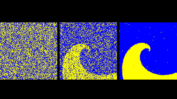
Convergence of a Monte Carlo Spiral - Hayden Sutton
|
These pictures show a Monte Carlo simulation of two particle types in a spiral made of two materials, progressing in time from left to right. The particles start disordered, moving randomly throughout the materials. As time progresses, more particles are created within their preferred materials, revealing the true shape of the structure. This illustrates the nature of the Monte Carlo technique; by using the chaotic but well-understood statistics of particle movement, the solution will be found regardless of the problem geometry, simply because the statistics will converge to that solution.
|
|
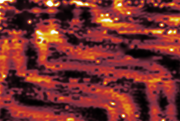
Gold Herringbones - Connor Vernachio
|
The above image is taken of atoms of a gold film, Au(111). It is taken from a scanning electron microscope, about 15 nm by 20 nm. Gold has a face-centered cubic structure, and the particular film is a 45 degree angle cut of that structure. As a result, there is a "herringbone" structure that appears, which are zig zagging atoms that are pushed 0.02nm above the surface of the material. The cut that makes the film creates tensile stress on the dangling atoms on the surface of the material, and they kink outward as if they are elastic.
|
|
|
|
Special thanks to the MSE graduate students who organized this event: Kim Pestovich, Sean Drewry and Sheryl Sanchez.
|
|
|
|
More photos on the Keffer research group site.
|
|
|